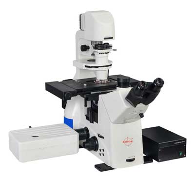
Confocal Microscopes
Confocal Microscopes RTC-7CON
Confocal microscope is a high-end product in Radical Optics microscope series. It is designed as an essential microscopy tool for laboratory scientific research, providing powerful and stable imaging capabilities and highly integrated motorization capabilities.
Efficient scanning head, detector and CVT motorized small hole, coupled with powerful optical system, provides fast, stable, high signal-to-noise (S/N) ratio confocal image.
Confocal Microscope provides a variety of motorized parts, including: motorized stage, motorized focusing, motorized nosepiece, motorized fluorescent carousel, motorized condenser and motorized brightness adjustment, operation mode allows physical button operation and software operation, and provides calling commands, which is convenient for users to control and develop by themselves.

Confocal Microscope provides a variety of motorized parts, including: motorized stage, motorized focusing, motorized nosepiece, motorized fluorescent carousel, motorized condenser and motorized brightness adjustment, operation mode allows physical button operation and software operation, and provides calling commands, which is convenient for users to control and develop by themselves.

- FEATURES
- SPECIFICATION
- - High resolution images can be generated with a single click operation, The software will automatically calculate size of the small hole according to objective numerical aperture, exposure value and scanning range, so as to obtain the image with the optimum signal-to noise(S/N) ratio.
- - At same time, noise reduction algorithm can remove the background noise in real time and improve image quality. Multi-channel images can be collected and synthesized simultaneously, which is convenient for customers to realize real-time observation of multiple stains.
- - By setting top position, bottom position and movement interval, the Confocal microscope motorized Z axis can realize automatic Z-Stack acquisitionand generate 3D model.
- - Providing various microscope motorized control interfaces: motorized objective carousel, motorized fluorescent filter unit, motorized condenser turntable.
- - Motorized stage control and motorized focusing mechanism could locate the Region of Interest (ROI) immediately through the software and record the position so that the user will be able to return to the recorded position quickly.
| Part Name | Part Code | Description |
|---|---|---|
| Optical System | RCiOS | NIS60 infinite optical system |
| Eyepiece | RCEP1025 | 10×(25), EP17.5mm, adjustable diopter-5~+5, interface ø30 |
| Viewing Head |
RCTST45 | Seidentopf trinocular tube, inclined at 45°, interpupilary distance 47-78mm, eyepiece interface ø30, fixed visibility: 1) eyepiece/camera switch (100/0, 50/50, 0/100); 2) visualization/turn off visualization /bertrand lens position adjustable |
| Nosepiece | RC-6N | Motorized sextuple nosepiece (expansion slot), M25×0.75 |
| Condenser | RABC | 6-Position motorized control N.A 0.55mm, W.D 26mm phase contrast (10/20, 40, 60 optional), DIC (10X/20X/40X) optional empty hole |
| Illumination | RC10WL | Transmitted kohler illumination, 10W LED illumination |
| Epi-Illumination: wide-field fiber illumination, 6-position motorized fluorescent carousel (B, G, U standard outfit), motorized fluorescent shutter | ||
| Mechnical Stage | RCMC | Motorized control moving range 130mm x100mm (325 mm x 144 mm) maximum speed: 25mm/s, resolution: 0.1µm - repeat accuracy:3µm. mechnical adjustable slice clamp |
| Focusing | RXF10 | Coaxial coarse and fine adjustment, stroke: focus up 7 down 2, coarse stroke 2mm per rotation, fine stroke 0.002mm per rotation, manual and motorized control, minimun stroke 0.01um under motivated control. |
| Objective | RC10 | 10X, NA 0.45, W.D. 4.0mm, cover glass thickness 0.17 |
| RC20 | 20X, NA 0.75, W.D. 1.1mm,cover glass thickness 0.17 | |
| RC60 | 60X, NA 1.42, W.D. 0.14mm, cover glass thickness 0.17, Oil | |
| RC100 | 100X, NA 1.45, W.D. 0.13mm,cover glass thickness 0.17, Oil | |
| Intermediate | RCI | Manual 1X,1.5X?Confocal switching |
| Output Port | RCOP | Splitting Ratio: Left:Eyepiece=100:0; Right:Eyepiece=100:0 |
| DIC Plate | RCDP | 10X,20X,40X Plate; Can be Inserted in Nosepiece Slot; Optional |
| Controller | RCC | Rocking Bar, Controller Box, USB Connection Cable |
| Laser Unit | RCLU | Laser 405 nm,488 nm,561 nm,640 nm |
| Detector | RCD | Wavelength: 400-750nm,Detector:4 PMT |
| Scanner |
RCS | Maximum Pixel Size: 4096 Scanning speed: 2fps(512X512), 18fps(256X256), 0.5fps(1024X1024), 0.12fps(2048X2048), 0.03fps (4096X4096) |
| Scan Mode | RCSM | X-Y, X-Y-Z, X-Y-T |
| Pinhole | RCPH | Hexagon shape, Continuouslv Variable Transmission(CVT) |
| Confocal | RCCF | Field number Square Inscribed in a ø18mm Circle |
| Image bit depth | RCIBD | 12 bits |
| Compatible Microscopes |
RCCM | Full Motorized Inverted Microscope |
INSTRUMENTATION SINCE 1975 © 2024 Radical Scientific Equipments Pvt Ltd, All rights reserved


