Focus: A comprehensive, application-focused selection guide designed to help lab directors, educators, and purchasing officers identify the ideal laboratory microscope. We balance visual and optical performance with mechanical ergonomics and workflow efficiency to optimize your investment.
Buyer's Guide
How to Choose the Right Laboratory Microscope for Research and Educational Labs
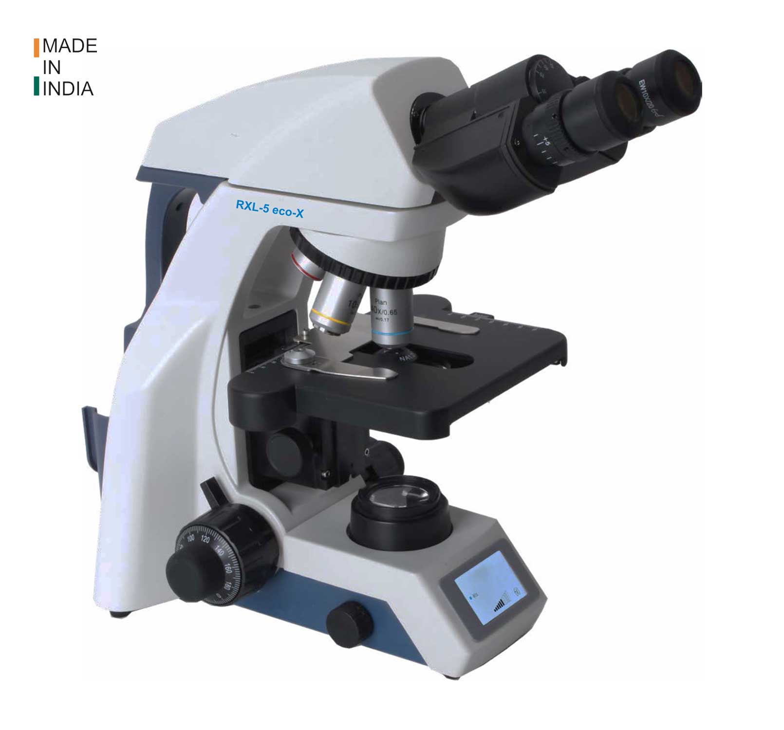
Why microscopes matter in laboratories
Microscopes are critical instruments across life sciences, pathology diagnosis, materials engineering, and classroom research. A high-quality laboratory microscope does more than amplify details; it maximizes resolving power, produces high-contrast images, ensures experimental repeatability, and speeds up diagnostics. In compliance-heavy settings like clinical pathology or GLP labs, precise instrument specs are vital for audit readiness and patient outcomes.
Types of laboratory microscopes
Choosing the correct class of instrument is essential to matching your imaging tasks. Below is an analytical overview of the primary categories, along with a comprehensive comparison matrix to help you determine which microscope is best for your laboratory.
Biological microscope (compound)
This is the standard upright or inverted configuration for slide-based cell biology and cytology labs. It supports transmitted light modes (brightfield, phase contrast, and DIC) and serves as the baseline microscope for pathology labs. Explore our tailored product configurations: Laboratory Clinical Microscopes.
Fluorescence microscope
Combining high-intensity excitation light sources and specialized interference emission filters, fluorescence microscopes detect specific molecular fluorophores. These are mandatory for immunoassays and cell assays. Discover our solutions: Fluorescent Microscopes.
Stereo microscope
Also known as dissecting microscopes, these provide a distinct three-dimensional stereoscopic view, wide field-of-view, and large working distances. They are vital for specimen prep, dissection, and industrial electronics inspection. View options: Stereo Microscopes.
Polarizing microscope
Featuring rotatable polarizing stages and analyzers, these instruments reveal structural birefringence and optical anisotropy in rock thin-sections, minerals, polymers, and crystals. Check our metallurgy range: Metallography Equipments.
Digital microscope
By eliminating eyepieces in favor of integrated CMOS sensors, digital microscopes enable real-time projection, precise multi-point measurements, and swift cloud-sharing of images. See digital camera and software options: Digital Imaging & Software and Microscope Cameras.
| Microscope Type | Primary Application | Magnification Range | Key Advantage |
|---|---|---|---|
| Biological (Compound) | Pathology, cell slides, cytology | 40x – 1000x | High Resolution |
| Fluorescence | Immunology, cell assays, FISH | 100x – 1000x | Molecular Specificity |
| Stereo (Dissecting) | Dissection, industrial QC, assembly | 5x – 100x | 3D Depth & Space |
| Polarizing | Geology, crystallography, polymers | 40x – 600x | Crystalline Analysis |
| Digital | Documentation, team streaming | 10x – 5000x | Easy Archival |
Applications by microscope type
Application requirements are the primary guide for microscope specifications. Here is how specialized research applications shape your choices:
- Pathology and Clinical Diagnosis: Prioritize ultra-flat optical field correction (Plan objectives) and standard high-contrast brightfield transmitted light options to examine cellular tissue abnormalities.
- Sample Handling & Dissection: Require stereoscopic optical paths to evaluate material depth, along with customizable arm-stands and external gooseneck fiber illumination.
- Molecular Research: Require fluorescence excitation led sources, high quantum-efficiency cameras, and specific filter cubes designed to block signal crosstalk.
- Metallurgy & Polymer Chemistry: Utilize reflected polarized light setups to observe surface grain boundaries, cracks, crystallization stages, and stress fatigue points.
Key factors to consider before purchasing
Choosing a microscope involves looking at it as an integrated imaging system rather than just a standalone magnifier. Always keep these key parameters in mind:
Optical Correction — Lenses are Decisive
Objectives determine the limit of what your microscope can resolve. Plan to spend a significant portion of your budget on optics:
- Achromat Lenses: Standard correction for biological slides, corrected for blue/red colors. Perfect for teaching.
- Plan Lenses: Corrected for spherical curvature to ensure the image remains crisp and in-focus from the center to the outermost edges. Required for pathology diagnosis.
- Apochromat Lenses: Corrected for three colors, offering peak resolution and chromatically perfect color replication. Essential for publishing and molecular imaging.
Digital Integration & Camera Optimization
Match the camera sensor size and pixel density to your objective lens's numerical aperture (NA). This uses the Nyquist sampling theorem to preserve details without causing optical empty magnification.
Technical Tip: Always prioritize purchasing the highest quality objectives and essential fluorescence filters first. Accessories like camera hardware, motorized stages, and advanced analysis software can easily be retrofitted later.
Educational vs research selection
Educational compound setups (colleges & secondary schools)
Prioritize mechanical durability: integrated stage lock stops to prevent slide breakage, fixed eyepiece tubes, robust led illumination, and locked components to withstand high student throughput. View options: Educational Microscopes.
Advanced research systems
Require advanced modularity: phase contrast and DIC sliders, high-aperture oil/water immersion objectives, high sensitivity cameras, and modular live-cell environmental control chambers.
Practical buying checklist
Use this six-step checklist to systematically guide your procurement team through the microscope evaluation process:
1
Define Sample Formats
Identify whether your slides or petri-wells require an upright or inverted microscope frame.
2
Specify Lens Correction
Match your budget to objective lens types: standard Achromat, flat-field Plan, or high-end Apochromats.
3
Optimize Camera Sampling
Ensure camera sensor pixel sizes perfectly align with objective NA to prevent empty magnification.
4
Confirm Light Sources
Select uniform LED transmitted lighting or stable, high-intensity fluorescent LED systems.
5
Secure Service Agreements
Choose manufacturers that supply localized service engineers, calibration, and replacement parts.
6
Verify with Demos
Request live sample imaging tests or on-site demonstrations to evaluate mechanical response.
GLP Lab Requirement: For diagnostic or clinical laboratories, verify that your scientific microscope supplier provides comprehensive IQ/OQ validation certificates to maintain quality standards.
Article Visuals Gallery
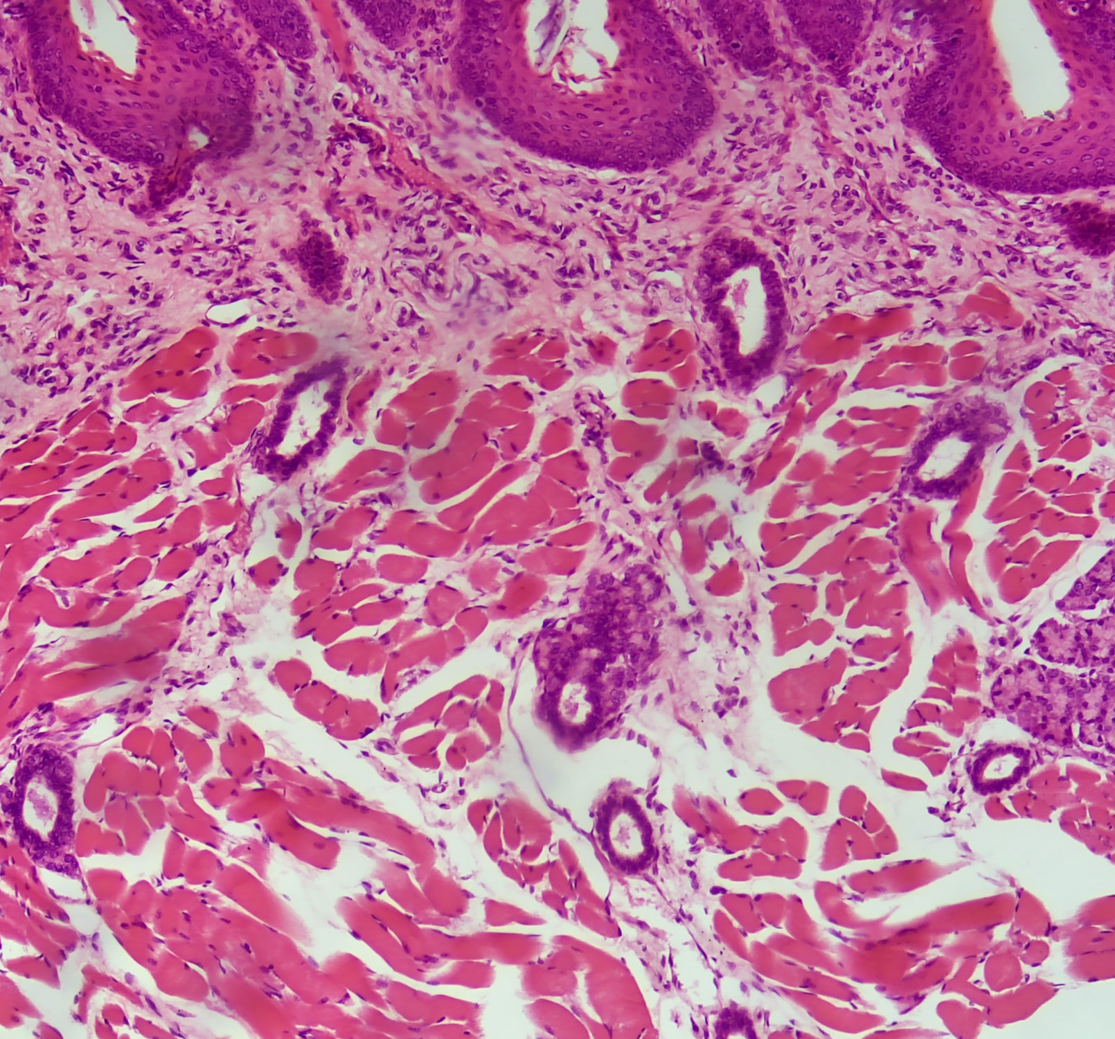
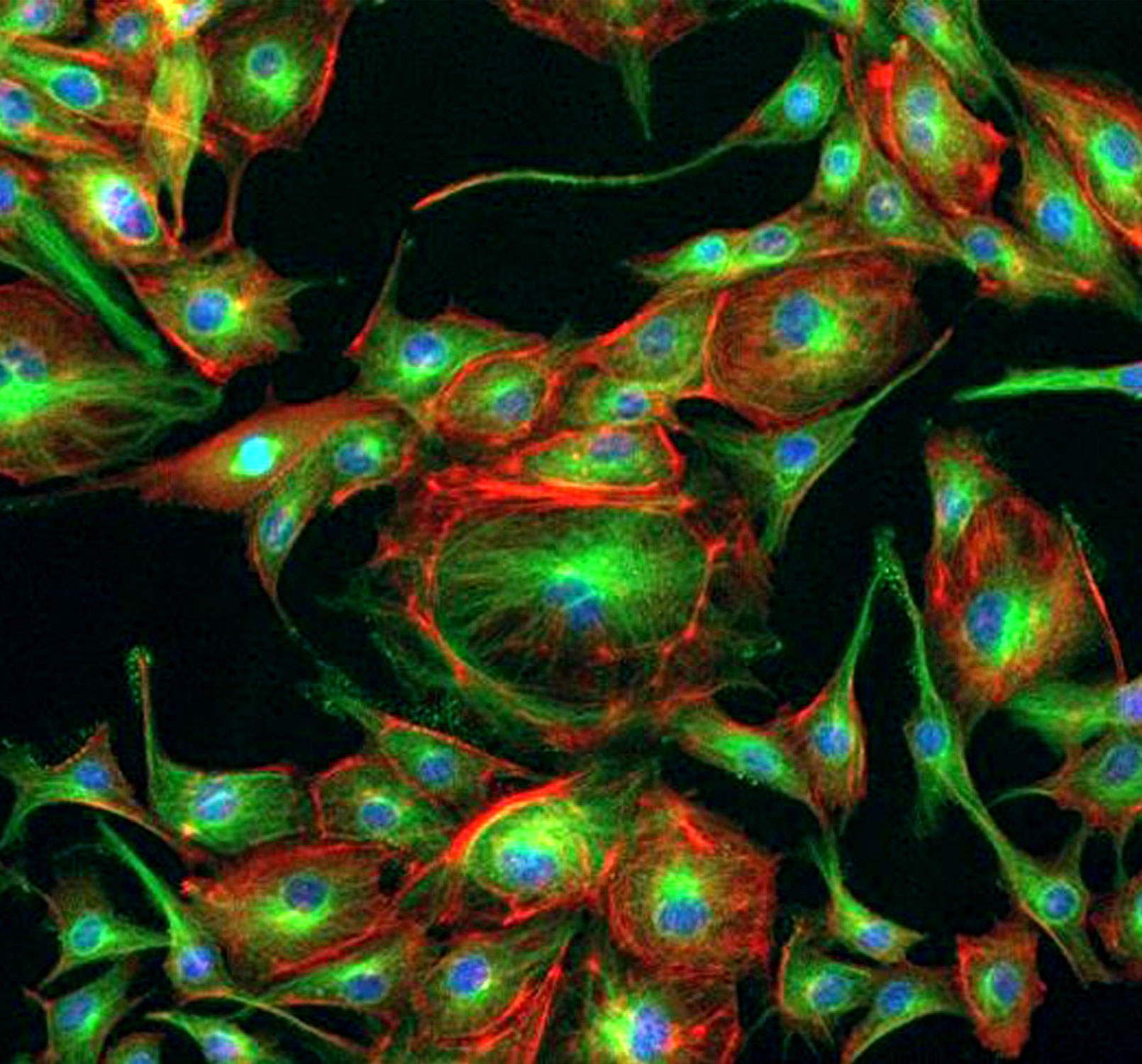
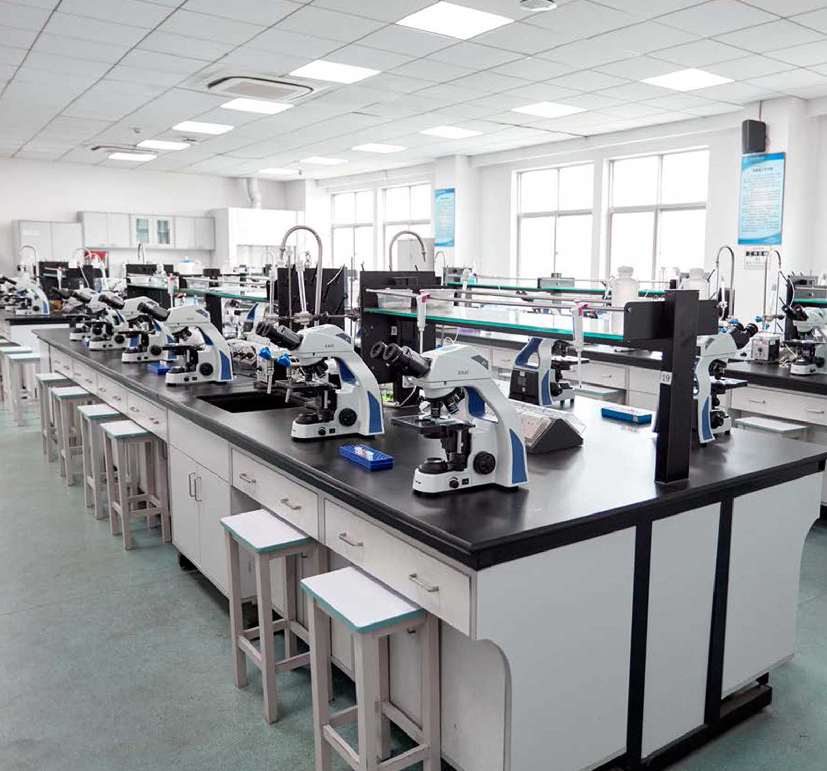
Reference microscope systems demonstrating ideal hardware configurations for pathology, education, and molecular diagnostics. Constrained layout frames ensure sharp images on all screens.
Frequently Asked Questions
What is the difference between a biological microscope and a stereo microscope?
A biological (compound) microscope uses transmitted light and high-magnification objectives to resolve cellular features. A stereo microscope provides lower magnification but gives true 3D depth, making it ideal for micro-dissection and component assembly.
Do pathology labs need a fluorescence microscope?
Most routine pathology work is brightfield-based. However, clinical diagnostics require fluorescence setups for specialized immunological assays, molecular pathology, and target-specific FISH techniques.
How do I match a camera to an objective?
Match camera pixel dimensions to the objective's optical resolving power, using Nyquist sampling guidelines. This ensures your imaging chip records all details resolved by your optics. Our support team can assist with exact calculations.