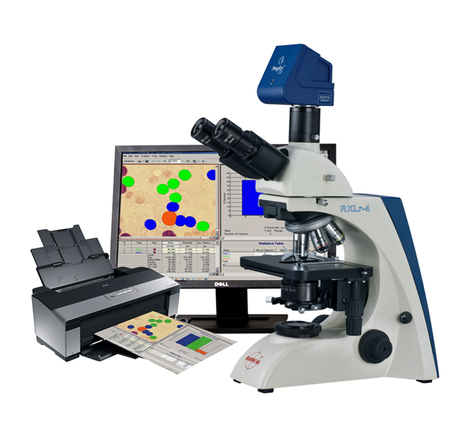
RI Morphology
Provides enhancement and measurement of the images. The images and data can be stored in album image database. RI(morphology can be used as research-purpose image analysis software. It also includes several pre-installed methodics (macros) for specific biomedical applications.
| RI (Morphology) | includes 6 pre-installed methodics (macros): |
|---|---|
| Measure | Objects' thresholding and measurement. |
| Count and Measure | Objects' thresholding, measurements, and automatic classification. |
| Volume Fraction | Thresholding the image phases. The thresholded phases are measured, and the area percentage of different phases is calculated. |
| Platelets Count | Thresholding the erythrocytes and platelets, their recognition and count. |
| Erythrocytes Sizing | Thresholding the erythrocytes, their measurement and classification by size |
| Nuclear-Cytoplasm Ratio (NCR) | Calculation of the area ratios (nucleus to cytoplasm). |
High image quality: Color interpolation, measured data based on EMVA 1288 guidelines, real time enhancements @ live image: sharpness, noise reduction, dynamic, white shading, colors balance, contrast saturation.
Versatility: WIN MAC Linux, Research Life – with free multi-fluorescence tool, Examine Elements – with free panorama & z-stacking tool, Evaluate Quality – with free measurements & annotation tools, Reveal Truths – get best color reproduction based on measurements and true color know how.
Ease of use: Connect the camera, launch software and start recording data, automatic, fast exposure control, live-image optimized: panorama, stitching, z-stacking, multi-fluorescence, image comparison (side by side) & video recording and live-image optimized: ocular view - adapt color impressions from ocular to the images.
Stability: Device configuration - to define multiple microscope settings, user profile – for excellent, reproducible image results and continuously and free software upgrades.
Fields of Application
Research Life - Life sciences: Medicine, pathology, hematology, cytology, genetics, biology and chemistry.
Evaluate Quality – Quality control: Grain analysis, welded seam testing and controlling manufacturing processes.
Examine Elements - Material science: Mineralogy and metallography-for use in determining structures, quantitative and qualitative sample analyses and documentation.
Reveal Truth -Forensics - Securing of evidence, document examination and forensic medicine.
Morphology software provides enhancement and measurement of the images acquired with a CCD/CMOS camera installed on the microscope. The analysis data are processed statistically. The images and data can be stored in Image Database. (Morphology) can be used as general-purpose image analysis software. It also includes several pre-installed methodics (macros) for specific biomedical applications.
Morphology main functions:Morphology includes 6 pre-installed methodics (macros):
Measure: Objects' thresholding and measurement.
Count and Measure: Objects' thresholding, measurements, and automatic classification.
Volume Fraction: Thresholding the image phases. The thresholded phases are measured, and the area percentage of different phases is calculated.
Platelets Count: Thresholding the erythrocytes and platelets, their recognition and count.
Erythrocytes Sizing: Thresholding the erythrocytes, their measurement and classification by size.
Nuclear-Cytoplasm Ratio (NCR) : Calculation of the area ratios (nucleus to cytoplasm).