How to Choose the Right Microtome for Consistent, Diagnosis-Ready Tissue Sections
By Radical Scientific — Applications TeamPublished: 16 July 2026
Focus: A practical guide for histotechnicians, pathologists, and lab procurement teams on matching specimen type, sectioning volume, and precision requirements to the correct microtome — covering rotary, cryostat, sliding, rocking, and fiber microtomes, with real specifications from Radical Scientific's manufacturing range.
A microtome is only as good as the sections it produces. Every histopathology lab has faced the same frustrating moment: a section comes off the blade torn, chattered, or unevenly thick, the slide goes into the bin, and a biopsy that cannot always be repeated has to be re-cut from the block. In frozen section work, that same delay happens while a surgical team is waiting in the operating theatre for a diagnosis. Both situations trace back to the same root cause more often than lab staff realise — the microtome in use does not match the specimen preparation method, the sectioning volume, or the precision the diagnosis actually requires.
This guide walks through how to diagnose that mismatch and select a microtome that eliminates it.
why tissue sections fail
Poor sectioning is rarely a skill problem alone — it is usually an equipment mismatch. The most common section failures and their underlying causes:
What you see on the slide
Underlying cause
Consequence
Uneven or variable thickness across the section
Feed mechanism precision too coarse for the tissue type, or worn feed components on an ageing microtome
Poor cell morphology, unreliable special stain results
Chatter marks / venetian-blind striping
Wrong knife angle or clamp rigidity for a dense or calcified specimen
Diagnostic detail obscured; section unusable
Sections tear or compress
Frozen specimen cut on a rotary microtome instead of a cryostat, or vice versa for paraffin blocks
Lost turnaround time in intra-operative diagnosis
Blade damage on the specimen face during the return stroke
Microtome without automatic specimen retraction
Irregular subsequent sections, shortened blade life
Bottlenecked sectioning during high specimen volume
Manual rotary microtome used at a throughput it was not built for
The fix starts with specimen preparation, not brand comparison. Before comparing models, identify how your specimens actually reach the blade — paraffin-embedded, frozen, celloidin-embedded, or industrial (fibre, rubber, plastic). This single factor eliminates most of the wrong-microtome mismatches described above.
Step 1 — Match the microtome to your specimen preparation method
This is the single most important decision in microtome selection. The specimen preparation method determines the entire microtome category — everything else is refinement within that category.
Specimen Preparation
Typical Use Case
Correct Microtome Type
Paraffin-embedded (routine histology)
Hospital pathology labs, diagnostic centres, research institutes
Step 2 — Weigh automation level against your sectioning volume
Once specimen type has fixed the category, automation level is what determines throughput, operator fatigue, and consistency at scale — and this is where a general guide reaches its limit. The right tier depends on your actual daily specimen count, staffing, and how much manual cranking your technicians can sustain across a shift without fatigue-related inconsistency creeping into sections.
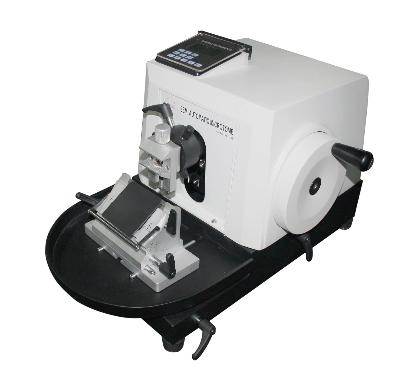
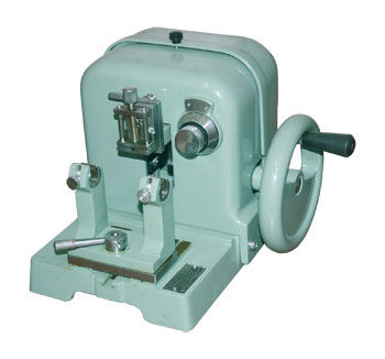
Hand-cranked feed with a fixed increment, a heavy cast-iron base, and a rotating knife clamp with lateral movement. The simplest, most economical tier — well suited to labs where sectioning is not the throughput bottleneck.
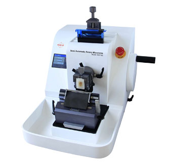
A stepper motor drives the feed with continuous, single-step, or programmed sectioning modes, controlled from a touch display. Motorised coarse feed removes the repetitive hand-cranking that fatigues technicians during long sectioning runs, and features like automatic specimen retraction protect the block face between cuts.
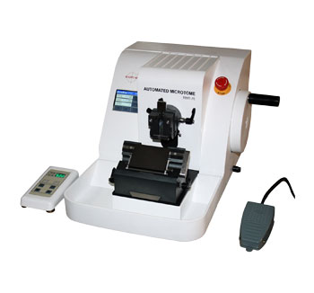
Runs fully automatic or manual from a single instrument, with foot switch and remote control operation. Built for the highest sectioning volumes, where manual cranking is no longer practical for an entire shift.
Our range covers all three tiers — from entry-level manual models through to fully automatic systems, each with different feed ranges and safety features suited to different daily volumes. Rather than guess which tier and model fits your lab, our expert engineers will match the exact microtome to your specimen count and staffing after a short conversation — talk to an expert engineer.
Step 3 — Confirm section thickness precision against your diagnostic requirement
Routine histopathology typically requires 3–5 micron sections for clear nuclear detail under the microscope. Confirm the feed increment of any microtome you are evaluating is fine enough to hit this range reliably, not just capable of it at the coarse end of its range.
A microtome that only offers 5-micron increments cannot reliably produce the 3-micron sections many special stains and immunohistochemistry protocols require. Verify the fine-end increment specifically, not just the overall range printed on the specification sheet — this is exactly the kind of detail worth confirming with our expert engineer against your specific staining and diagnostic protocol before you commit to a model.
Special case — frozen section and intra-operative diagnosis
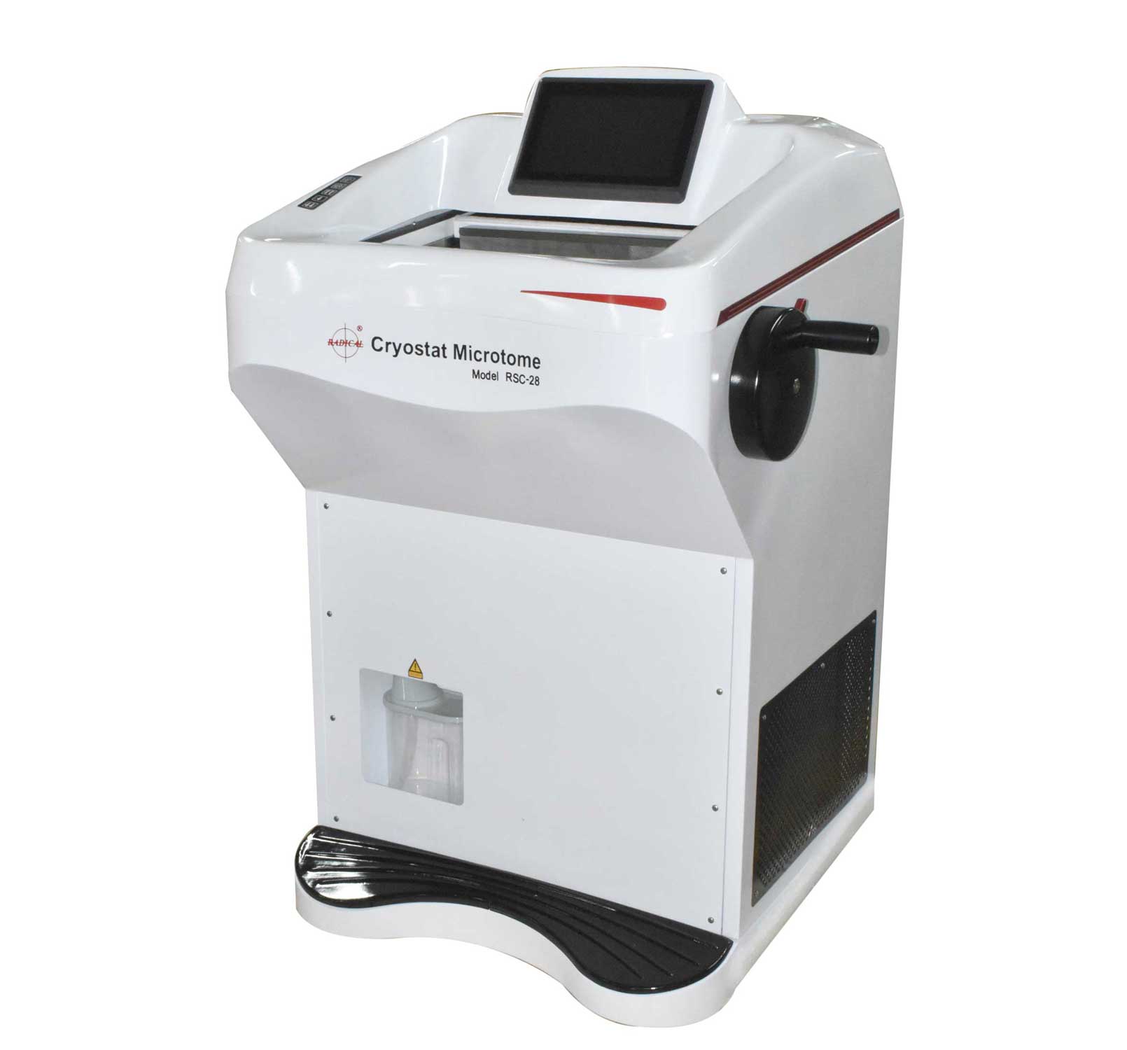
Frozen section diagnosis is the highest-pressure application a microtome will ever face — a surgeon and patient are waiting on the outcome, typically within a 20-minute turnaround window. A rotary microtome designed for paraffin blocks cannot be substituted here; the specimen must be cut inside a refrigerated chamber immediately after freezing.
The cryostat microtome is purpose-built for this: a freeze chamber running from −10°C to −40°C, dual German compressors, UVC sterilisation, a heated sliding window to prevent condensation fogging during use, and a 17-cassette specimen shelf so multiple cases can be staged without warming the chamber. Section thickness runs from 1–100 micron with 1-micron increments at the fine end, matching routine paraffin precision despite the frozen state of the tissue.
Special case — industrial fibre and material testing
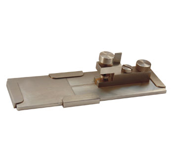
Fibre microtome for cross-section analysis
Not every microtome application is medical. Textile, rubber, and plastics laboratories use a fibre microtome to produce cross-sections for diameter and fibre-type identification under a microscope, to recognised material testing standards including IS 744, ASTM D2130, BS 2043, DIN 53811, ISO 137, and IWTO 8-6. This is a materials quality-control instrument, not a diagnostic one — but the same principle applies: the microtome must match the specimen and the standard being tested against, not be a general-purpose substitute.
Complete buying checklist
✓ Specimen Type
Paraffin-embedded → Rotary microtome
Frozen / intra-operative → Cryostat
Large / celloidin → Sliding microtome
Industrial fibre → Fibre microtome
✓ Volume & Automation
Average daily specimen count noted
Staffing and shift pattern confirmed
Automation tier to be matched by our expert engineers
✓ Precision
Fine-end increment confirmed (not just range)
3–5 µm achievable for routine histology
Specimen retraction to protect block face
✓ Safety & Support
Automatic feed disengage / blade guard
Manufacturer with service support in your region
Accessories and blade supply confirmed
Microtomes from Radical Scientific
Radical Scientific has manufactured histopathology sectioning equipment since 1975. The current range covers every specimen type and automation level discussed in this guide, from entry-level manual rotary microtomes to fully automatic and cryostat systems.
View the complete range on the microtomes gallery page, or the full histopathology equipment range including tissue processors, embedding stations, and slide staining machines. For full specifications, drawings, and pricing of any model, contact our expert engineers directly — we will recommend the right instrument for your specimen type and sectioning volume.
Frequently asked questions
What is the difference between a rotary microtome and a cryostat?
A rotary microtome sections paraffin-embedded tissue at room temperature. A cryostat is a rotary microtome housed inside a refrigerated chamber, used to section fresh frozen tissue — most commonly for intra-operative diagnosis during surgery, where results are needed within minutes.
What section thickness does routine histopathology require?
Routine histology sections are typically cut at 3–5 microns for clear nuclear and cellular detail under the microscope. Confirm the fine-end feed increment of any microtome — not just its overall range — supports this thickness reliably.
Should I choose a manual or automatic microtome?
This depends on your daily specimen count, staffing, and shift pattern — factors that vary enough between labs that a general rule can be misleading. Our expert engineers reviews your actual sectioning volume and recommends the right automation tier for your lab specifically; contact us for a direct recommendation.
Why do my sections tear or show chatter marks?
Torn sections usually indicate a frozen specimen cut on equipment not designed for it, or a feed mechanism too coarse for the tissue. Chatter marks typically come from knife angle or clamp rigidity mismatched to a dense or calcified specimen. Both are equipment-matching issues, addressed by selecting the correct microtome type and confirming its feed precision before purchase.
What is a fibre microtome used for?
A fibre microtome produces cross-sections of textile, rubber, and plastic fibres for diameter measurement and fibre-type identification under a microscope, to material testing standards such as IS 744, ASTM D2130, BS 2043, DIN 53811, and ISO 137. It is used in industrial material testing labs, not diagnostic pathology.
How much does a microtome cost in India?
Contact Radical Scientific at 1800-1233-229 or info@radicalscientific.com for current pricing and model recommendations based on your specimen type, sectioning volume, and precision requirements.