Phase Contrast Microscope: Complete Guide for Research and Clinical Labs
By Radical Scientific — Applications TeamPublished: 6 June 2026
Focus: A complete technical and application guide to phase contrast microscopy — covering the underlying optical principle, practical laboratory uses, a direct comparison with brightfield and DIC imaging, and a step-by-step selection framework to help you choose the right phase contrast microscope for your research or clinical workflow.
Phase contrast microscopy is one of the most significant advances in optical microscopy of the twentieth century. It allows researchers to examine living, unstained biological specimens in high contrast without the chemical fixation or staining that would kill the very cells under observation. If your lab works with live cells, tissue culture, spermatozoa analysis, or any transparent biological specimens, a phase contrast microscope is not optional — it is essential.
What is phase contrast microscopy?
Phase contrast microscopy is an optical imaging technique that converts differences in the refractive index of a specimen into corresponding differences in light intensity — producing high-contrast images of transparent structures that would appear nearly invisible under standard brightfield illumination.
The technique was invented by Dutch physicist Frits Zernike in 1934, for which he was awarded the Nobel Prize in Physics in 1953. The core optical principle rests on the behavior of light passing through a specimen with varying optical path lengths.
When light passes through a transparent biological structure such as a cell membrane, organelle, or nucleus, it is not absorbed — it is phase-shifted. That is, the wave of light exits the specimen slightly behind the surrounding wave that passed through the medium unimpeded. This phase difference of approximately one-quarter wavelength is invisible to the human eye and to standard camera sensors. The phase contrast optical system converts this invisible phase difference into visible amplitude (brightness) contrast through two engineered optical elements:
Annular diaphragm (condenser annulus): Positioned in the front focal plane of the condenser, this ring-shaped aperture illuminates the specimen with a hollow cone of light. It replaces the standard circular condenser aperture.
Phase plate (phase ring in the objective): Located in the rear focal plane of the objective lens, this plate contains a phase-advancing or phase-retarding ring that aligns precisely with the diffracted light from the annular diaphragm. It retards the direct (undiffracted) light by one-quarter wavelength, allowing constructive or destructive interference with the diffracted light from the specimen.
The result is an image where denser cellular structures appear darker against a bright background (positive phase contrast) or brighter against a dark background (negative phase contrast), depending on the phase plate configuration. Either way, the structural detail of an unstained, living cell becomes clearly visible.
Phase contrast vs brightfield vs DIC — which does your lab need?
Understanding the difference between these three transmitted light imaging modes is critical before specifying or purchasing a biological microscope. Each mode serves a distinct purpose, and selecting the wrong one for your application will compromise image quality and experimental outcomes.
Feature
Brightfield
Phase Contrast
DIC (Nomarski)
Specimen type
Stained, fixed tissue sections
Live, unstained transparent cells
Live cells, surface topology
Contrast mechanism
Light absorption by stain
Refractive index difference
Optical path gradient
Specimen preparation
Fixation and staining required
None required
None required
Live cell imaging
Not suitable
Ideal
Good
3D relief appearance
Flat
Moderate halo effect
Strong pseudo-3D
Halo artefact
None
Present at edges
Minimal
Cost / complexity
Low
Moderate
High (prism required)
Typical use case
Pathology, histology H&E sections
Cell culture, IVF, microbiology
Advanced cell biology research
Key Insight: For the vast majority of live-cell biology, tissue culture, and microbiology labs, phase contrast delivers the best balance of image quality, specimen viability, and instrument cost. DIC produces superior optical sectioning and pseudo-3D rendering but requires additional prism components and significantly higher investment. For pathology labs working with stained H&E sections, brightfield remains the standard.
Phase contrast microscope applications — where it is indispensable
The phase contrast microscope has become the standard instrument across a wide range of research and clinical disciplines. Below are the primary application areas where no other imaging mode performs as effectively.
Cell culture and tissue culture monitoring
Routine monitoring of mammalian cell cultures — including HeLa, CHO, HEK293, and primary cell lines — requires daily visual assessment of cell morphology, confluency, and health status. Phase contrast is uniquely suited to this task because it delivers clear images of unstained adherent and suspension cells without removing the culture from its growth environment. Researchers can assess cell shape, nuclear visibility, vacuolization, and apoptotic changes in real time.
For tissue culture labs, an inverted phase contrast microscope — where the objective is positioned below the stage — is the standard configuration. This allows direct visualization through the transparent base of standard culture flasks (T25, T75, T175) and multi-well plates without removing the lid or disturbing the sterile environment.
Assisted reproductive technology (ART) and IVF
Phase contrast microscopy is the primary imaging modality in IVF laboratories for spermatozoa morphology assessment, oocyte grading, embryo evaluation (at cleavage and blastocyst stages), and polar body observation. The ability to evaluate living gametes and embryos without fixation or fluorescent labeling makes phase contrast indispensable in this application. Inverted phase contrast microscopes with heated stages are the standard IVF lab instrument.
Microbiology and bacteriology
Bacteria, protozoa, and other microorganisms are transparent under standard brightfield illumination and require Gram staining or other procedures before they become visible. Phase contrast eliminates the need for staining in many screening applications, allowing direct observation of microbial motility, morphology, and division in their natural state. This is particularly valuable in environmental microbiology and water quality testing where rapid field assessment is needed.
Haematology and blood analysis
While most routine haematology uses stained blood smears, phase contrast is invaluable for reticulocyte analysis, platelet aggregation studies, and red blood cell deformability measurements. The technique allows assessment of cell membrane integrity and cytoskeletal structure without the morphological artefacts introduced by fixatives and stains.
Quality control in pharmaceutical and biotech manufacturing
Bioprocessing facilities use phase contrast microscopy to monitor cell viability, detect mycoplasma contamination in cell cultures, and assess particulate matter in bioreactor samples. The speed of phase contrast — no staining preparation required — makes it well suited to in-process quality checks on production timelines.
Key specifications to evaluate when choosing a phase contrast microscope
A phase contrast microscope is a complete optical system, not simply a standard biological microscope with an accessory add-on. The phase contrast components — annular rings in the condenser and phase plates inside the objectives — must be precisely matched and optically aligned. Here is what to evaluate before purchasing.
Phase contrast objective lenses
Phase contrast objectives are designated by a Ph code indicating the matched condenser ring size: Ph1 (low NA condenser ring, for 10x objectives), Ph2 (medium ring, for 20x and 40x objectives), and Ph3 (large ring, for 100x oil immersion). Your microscope system must include the correct matched condenser annuli for each phase objective you intend to use.
100x Ph3 (oil immersion): Bacterial motility, fine subcellular detail at maximum resolution
For Plan objectives (flat-field corrected), the designation becomes Plan Ph1, Plan Ph2, etc. Plan objectives are essential for any documentation or imaging application where edge-to-edge sharpness matters.
Condenser design and NA
The condenser numerical aperture must match or exceed the highest objective NA in your phase contrast set. For phase contrast specifically, a turret condenser with individually selectable brightfield, Ph1, Ph2, and Ph3 positions offers the most practical flexibility. Swing-in Bertrand lenses for alignment verification are a useful addition for research-grade systems.
Upright vs inverted configuration
This is the single most important configuration decision and is dictated entirely by your specimen container:
Upright phase contrast microscope: Standard slides, Petri dishes (open), smear preparations, haematology samples. Objectives above the stage, condenser below.
Inverted phase contrast microscope: Culture flasks, multi-well plates, culture dishes, closed vessels. Objectives below the stage looking upward through the vessel base. Essential for cell culture and IVF labs.
Illumination system
Modern phase contrast microscopes use LED transmitted illumination rather than tungsten halogen lamps. LED systems offer critical advantages: instant-on stability (no warm-up drift that shifts phase ring alignment), longer life span, lower heat output (important for live-cell applications), and adjustable color temperature for optimal phase contrast rendering at each magnification.
Digital camera integration
Phase contrast images require cameras with adequate sensitivity and pixel resolution to capture the fine contrast gradients produced by the phase plate. For documentation and morphological grading (IVF, cell culture QC), a 5–12 megapixel CMOS camera with good low-light sensitivity is the standard. For time-lapse cell imaging, ensure the camera software supports triggered acquisition and multi-position stage scanning. See our range of compatible systems: Microscope Cameras and Digital Imaging & Software.
Configuration Tip: If your lab anticipates future expansion into fluorescence imaging, select a phase contrast biological microscope with an epi-fluorescence port. Adding fluorescence capability to a well-specified upright or inverted phase contrast frame costs significantly less than purchasing a separate fluorescence system. Plan for modularity from the outset.
Phase contrast microscope selection checklist
Use this structured checklist to define your system requirements before approaching a supplier for a quotation. Having precise specifications reduces the risk of purchasing an underpowered or incorrectly configured instrument.
1
Define Specimen Containers
Identify your primary vessel: slides → upright; culture flasks or well plates → inverted. This determines the fundamental frame configuration.
2
Specify Magnification Set
List the magnifications you require (10x, 20x, 40x, 100x) and confirm the matched Ph condenser rings are included for each objective.
3
Choose Objective Correction
Select Plan objectives for even flat-field imaging across the full frame. Achromat Ph objectives are suitable for teaching; Plan Apochromats for publication-grade imaging.
4
Confirm LED Illumination
Specify LED transmitted illumination for instant stability. For live-cell work, verify the illumination intensity is adjustable to minimize phototoxic effects.
5
Plan Camera & Software
Match camera pixel size to the objective NA. For time-lapse, confirm multi-position and triggered acquisition capability in the imaging software.
6
Plan Future Modularity
If fluorescence or DIC may be needed later, confirm the frame accepts epi-fluorescence modules and DIC prism sliders without a full system replacement.
IVF and GLP Labs: For assisted reproduction or regulated analytical laboratories, confirm that your phase contrast microscope supplier can provide IQ/OQ validation documentation and calibrated service agreements. These are mandatory requirements for NABL accreditation and international IVF clinic standards.
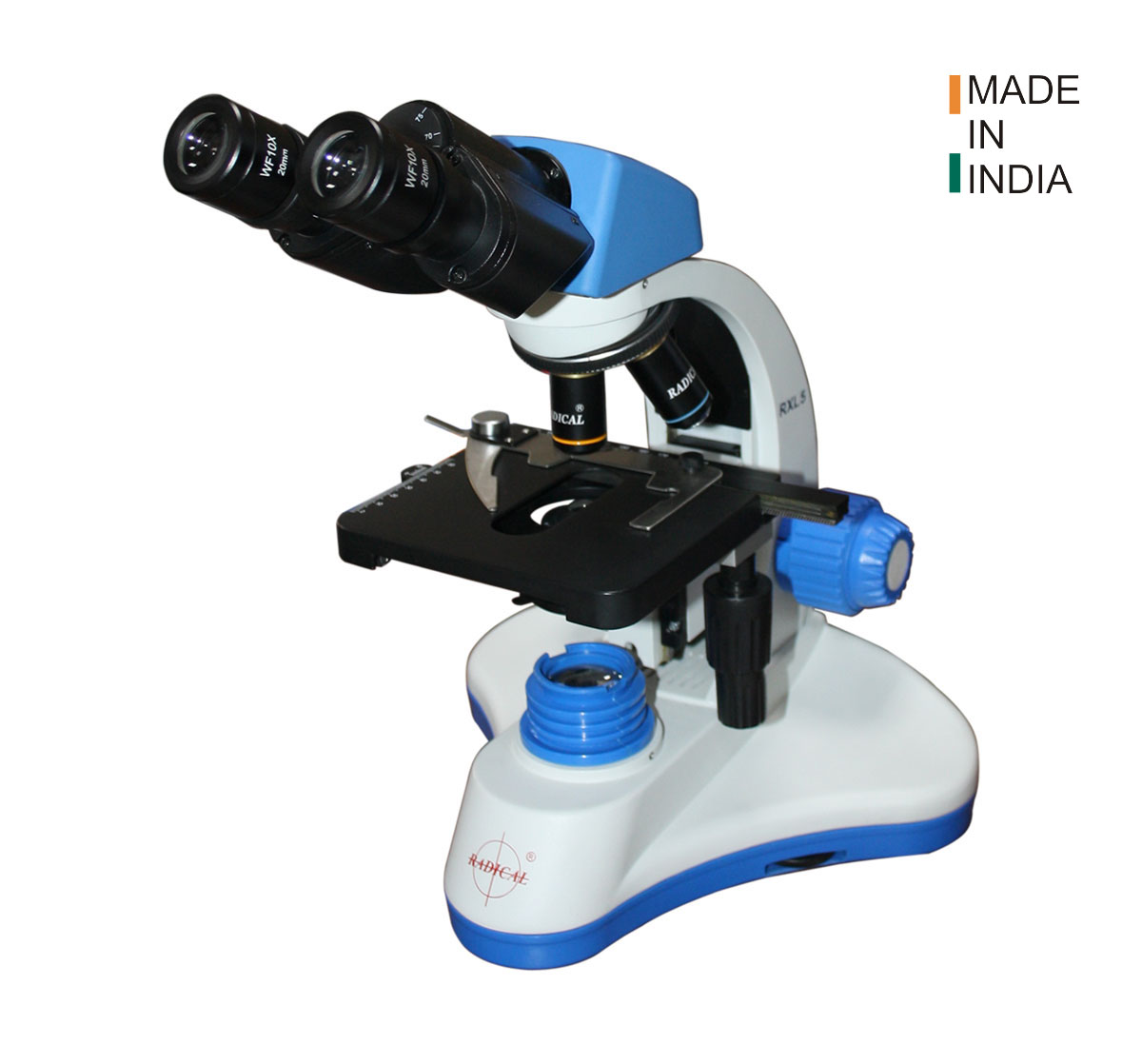
Phase contrast microscopes from Radical Scientific
Radical Scientific has manufactured biological microscopes with phase contrast capability since 1975. Our upright and inverted biological microscope ranges are engineered for demanding research and clinical workflows, with full optical compatibility across Ph1, Ph2, and Ph3 phase contrast systems.
Our laboratory and clinical microscopes support phase contrast as a standard or optional configuration across a range of frames — from robust educational systems for university labs to high-specification research microscopes with modular fluorescence and DIC expansion paths. All systems are supported by localized service engineers across India, with calibration and spare parts availability.
What is the difference between phase contrast and brightfield microscopy?
Brightfield microscopy requires specimens to absorb light — meaning they must be stained or naturally pigmented to produce visible contrast. Phase contrast microscopy converts differences in refractive index into brightness differences, making transparent, living, unstained cells visible without any specimen preparation. Phase contrast is therefore the standard imaging mode for live cell biology, while brightfield is standard for fixed and stained histology sections.
Can a standard biological microscope be converted to phase contrast?
Only if the microscope frame and condenser were originally designed to accept phase contrast components. Phase contrast requires a turret condenser with annular ring positions that precisely match the phase rings built into the objectives. A standard brightfield condenser and standard objectives cannot be modified for phase contrast. When purchasing a biological microscope with any anticipated need for phase contrast imaging, always confirm phase contrast compatibility at the time of purchase.
What is the halo artefact in phase contrast images?
The halo is a characteristic optical artefact of phase contrast microscopy — a bright rim of light that appears around the edges of structures in positive phase contrast images. It results from diffracted light from the specimen edge being partially transmitted by the phase plate alongside the direct beam. For most cell biology applications, the halo does not interfere with interpretation. For applications where it is problematic, apodized phase contrast objectives (which incorporate an additional neutral density ring) can significantly reduce halo intensity.
Is an inverted microscope required for tissue culture and IVF labs?
Yes, in almost all practical cases. Standard culture flasks (T-flasks), multi-well plates, and culture dishes are designed with optically transparent bases specifically for inverted microscopy. An inverted phase contrast microscope positions the objectives beneath the stage and illumination above, allowing the beam to pass through the vessel base and into the culture. An upright microscope cannot image through the opaque walls or lids of standard culture vessels. Inverted phase contrast configuration is the established standard for tissue culture, IVF, and bioprocessing labs.
What magnification is best for cell culture monitoring?
For routine culture monitoring — checking confluency, gross morphology, and contamination — a 10x Ph1 objective provides sufficient field of view for rapid assessment of the entire culture. A 20x Ph2 objective is the standard working magnification for cell morphology evaluation. A 40x Ph2 objective is used for detailed examination of individual cells, organelles, and mitotic figures. For bacterial cultures or fine subcellular detail, 100x Ph3 oil immersion is required. Most cell culture labs routinely work between 10x and 40x phase contrast.
What is the price of a phase contrast microscope in India?
Phase contrast microscope pricing in India varies significantly based on configuration. An entry-level upright phase contrast biological microscope suitable for a teaching or basic research lab typically starts from ₹35,000–₹60,000. A research-grade upright system with Plan phase contrast objectives, trinocular head, and LED illumination ranges from ₹80,000–₹1,80,000. Inverted phase contrast microscopes for cell culture and IVF labs, which require a larger and more complex frame, begin from approximately ₹1,50,000 for standard configurations and increase with motorized stages, camera systems, and environmental control accessories. Contact our sales team for a configuration-specific quotation: Request a Quote.