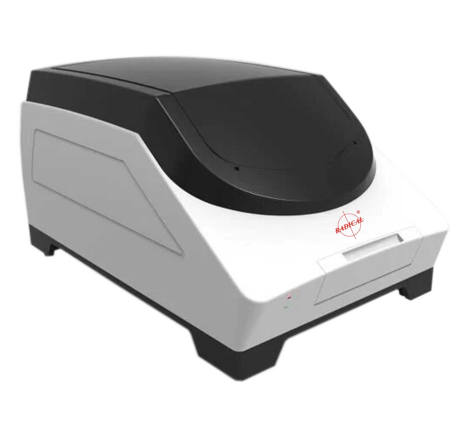
RDSS-1
The digital slide scanning range offers exceptional image quality, speed and reliability for whole slide imaging, making Nx Series scanners the optimal choice for healthcare and research professionals.
Unique Magnetic Absorption Type Glass Plate
Ensure accurate positioning of glass plate; Avoid repeated friction during loading and unloading, ensure smooth loading , and prevent sliders from falling and breaking during operation.
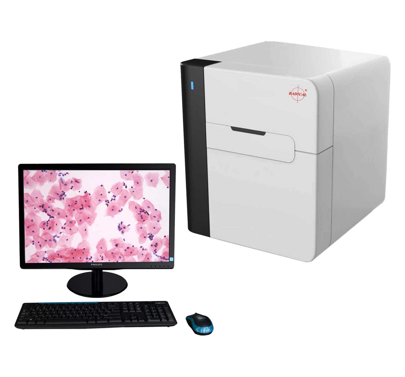
Scanning Software Features
- Real-time autofocus
- Real-time accurate positioning to view the scanned slide, equivalent to the function of microscope observation.
- Display image current position parameters and organization information parameters in real time
- When there are multiple pieces of tissue on the same slide, the software can intelligently identify them, scan them independently, and fuse them into a single digital slice
Adopt Strong and Weak Electric Sectionalized Control to Ensure the Safety of Personnel and Instrument
High Speed and Precision XY/Z Three-dimensional Platform System
-Imported maglev linear motor: Speed 3.2m/s, acceleration 8g
-High precision full closed loop drive control system, the X/Y axis motion resolution is 100nm, and the Z-axis motion resolution is 50nm
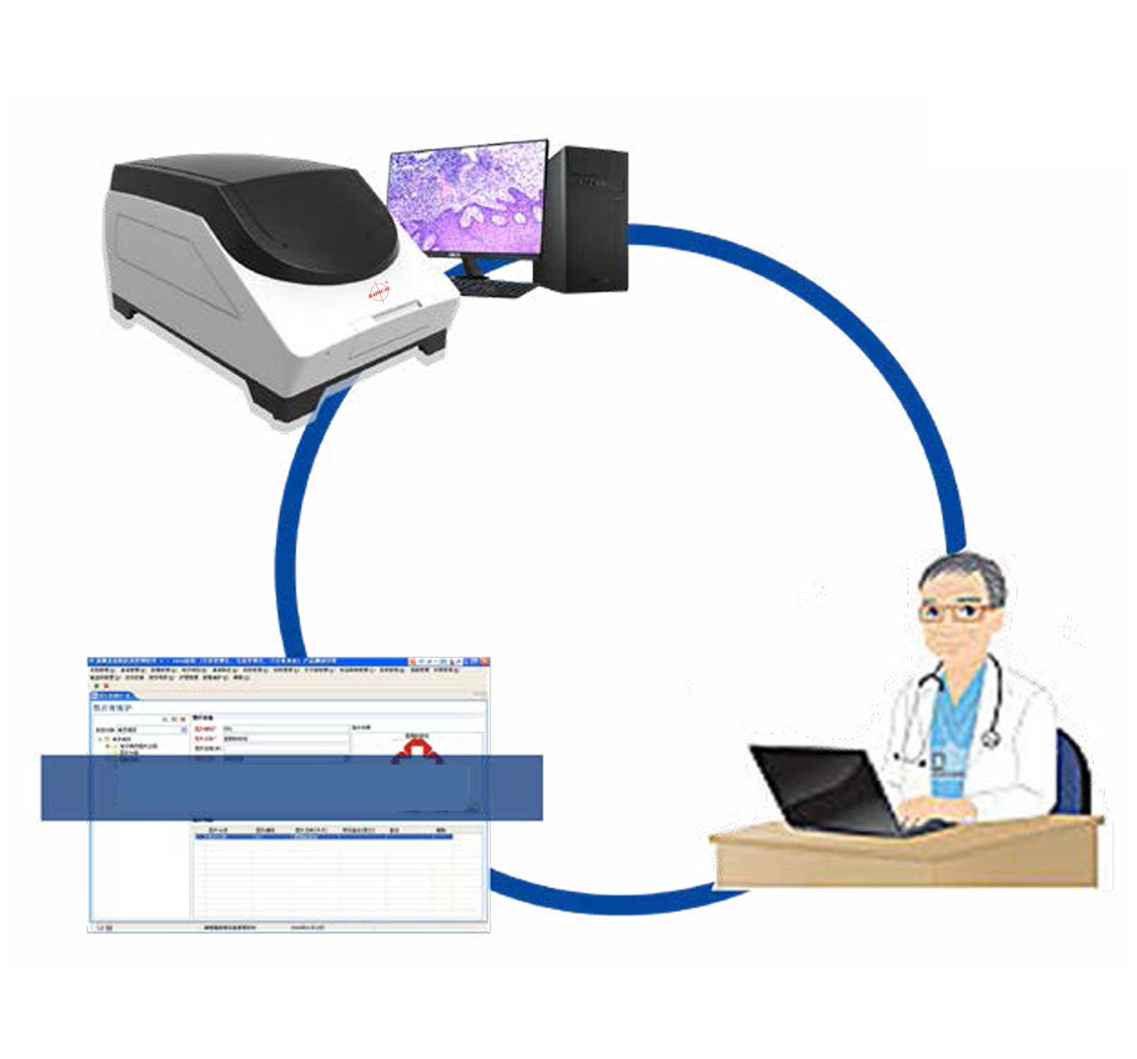
Browser Software
- With slice library management function.
- Color distinguish slice browsing track.
- Tag ROI with different shapes, colors, and line widths, and can export tag information to EXCEL files. Regional screenshots of 2300dpi and full screenshots can be made to meet the publishing needs of periodicals and books.
| Model | RDSS-1 | RDSS-FL |
|---|---|---|
| Slide Tray | 1-6 Slides Capacity with auto slides loader | 1-5 Slides Capacity with auto slides loader |
| Slide Dimensions | Size: 26X76mm ± 1mm tolerance on both side Thickness: 0.8-1.2mm, including coverslip |
Thickness: 0.8-1.2mm, size: 26X76mm |
| Objectives | 20x plan apochromatic objective, N.A. = 0.75 | 20x plan apochromatic objective, N.A. = 0.75 |
| Light Source | Customized LED Professional Light Source | |
| Magnification | 20x or/& 40x | 2x, 20x & 40x |
| Slide Identification | 1D/2D Bar code/QR code indetification/ manual | |
| Scanning platform | Upscale maglev linear motor. High precision full closed loop drive control system | |
| Scanning Resolution | ≤ 0.48μm/pixel(20X) ≤ 0.24μm/pixel(40X) |
≤ 0.67μm/pixel(2X) ≤ 0.67μm/pixel(20X) ≤ 0.45μm/pixel(40X) |
| Scanning Speed | Scanning area 15 mm × 15 mm, 20× Scanning time ≤ 40 seconds 40×(≤ 0.24μm/pixel) Scanning time ≤ 120 seconds |
2X 1 min/1 slide (26mmX76mm) Scanning area 15 mm × 15 mm, 20× Scanning time ≤ 40 seconds |
| Scanning Magnification | 40x | |
| Optical System | Infinity & Semi-Apochromatic corrected | |
| Focusing Technique | Real-time autofocus, provide a variety of scanning modes, to meet the scanning needs of various different sections: standard scanning, high-precision scanning, etc. Automatic identify the tissue location setting scan area, automatically skip the blank area, and also manually define the scan area. Capacity to scan uneven, folded, thick, broken tissue sections of Histology and cytology & also slides with fresh/wet cover slips, and/or protruding cover slips. | |
| Conversion | Glass slides into high quality digital scans | |
| Camera | Inbuilt Camera with resolution 7MP, Sensor Size: 2/3” to scan slides at resolution | |
| Professional Image Scanning, Management and Browsing Software | Batch scanning:
|
|
| Advantages |
|
|
| Fluorescence Turret | No | 5 position |
| Repeat positioning accuracy | ≤ 0.1um | |
| Scanning mode | Brightfield scanning | Brightfield or/& Fluorescence scanning |
| Illumination System | LED with fly-eye lens | |
| Field of vision | 1.3mm | |
| Software |
|
|
| NO | The fluorescent motorized turret B filter assembly G filter assembly U filter assembly V filter assembly R filter assembly |
|
| Weight | 25kg. | |
| The Depth of Field Fusion | Yes, upto 15 levels | |
| Barcode Support | Yes | |
| Computer | Processor: Intel i7, Memory: 16G, Hard Disk: 2TB, Monitor: 24" | |
| Temperature | Operating Temp.: upto 40°C, Storage Temp.: upto 60°C | |
| Power Supply | 90V-240V/50Hz AC (SMPS) with suitable UPS (Minimum 10 minutes backup) | |